Identification of Red & White Blood Cells
Blood is a liquid connective tissue.It is composed of a variety of cells circulating in a fluid, plasma. We are not interested in plasma in this lesson, only in the cells, both white and red blood cells. Blood cells are in three functional classes: red blood cells (erythrocytes), white blood cells (leukocytes) and platelets (thrombocytes). All three are formed in the bone marrow but have vastly different appearances and functions.
Red Blood Cells (Erythrocytes)
Red blood cells (RBCs) are the most numerous cell type in the blood (4.8-5.4 million RBCs/mL of blood). The cells are modified structurally to carry oxygen. The cells are biconcave disks approximately 8 µm in diameter (a doughnut without a hole) with no nucleus or metabolic machinery. The absence of cellular organelles allows the internal space of the cell to be available for O2carrying. The interior of the RBC is filled with Hemoglobin (Hgb), a protein that functions primarily to carry O2. A typical RBC has approximately 280 million Hgb molecules in it due to the lack of cellular organelles.
White Blood Cells (Leukocytes)
White blood cells are much less numerous than RBCs (5,000 – 10,000 WBCs/µL blood) with a RBC/WBC ratio of approximately 700:1. WBCs work to protect the body from infection. WBCs are divided into two main groups based on cytoplasmic appearance: agranular leukocytes (lymphocytes and monocytes that have relatively clear cytoplasm) and granular leukocytes (neutrophils, eosinophils, and basophils whose cytoplasm is filled with granules.
Granular Leukocytes
- NeutrophilsThe most abundant granulocyte, and most abundant WBC, is the neutrophil comprising 60-70% of all WBCs.They are approximately 10-12 µm diameter with very fine, pale lilac granules in the cytoplasm. The nucleus has from 2-5 finely connected nuclear lobes that are rarely uniform in size. The multiple lobed nucleus and lightly stained cytoplasm are the most identifiable characteristics of the this cell. Neutrophils respond rapidly by chemotaxis to bacterial damage. They then phagocytize pathogens and release lysozymes, strong oxidants and defensins to help fight the infection.
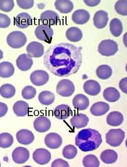
- EosinophilsEosinophils represent only 2-4% of all WBCs. They are similar in size to neutrophils, 10-12 µm diameter, with red/orange, large uniform granules, that do not block the nucleus. The nucleus usually has 2 but may have 3 connected nuclear lobes that tend to be more rounded and uniform than found in neutrophils. The red/orange color and more uniformly shaped lobed nucleus are the keys to identifying this cell. Eosinophils combat histamine in allergic responses, phagocytize antigen-antibody complexes and destroy some parasitic worms.
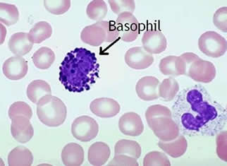
- BasophilsThe least common WBC is the basophil comprising only 0.5-1% of all WBCs. Basophils are 8-10 µm diameter with large blue-black round granules in the cytoplasm that block the bilobed nucleus. The dark granules are the most easily identifiable characteristic of this cell. Basophils exit capillaries and enter tissue fluids where they release heparin, histamine and serotonin during hypersensitivity (allergic) reactions to stimulate inflammation.
Agranular Leukocytes
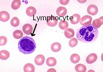
- LymphocytesLymphocytes make up 20-25% of all WBCs. These cells vary in size with small lymphocytes being 6-9 µm and large lymphocytes 10-14 µm in diameter. The nucleus stains dark and is round or slightly indented with the cytoplasm appearing as a rim around the nucleus. The round, uniform nucleus and small amount of cytoplasm surrounding it are the best identifying characteristics for this cell. Lymphocytes are involved in the specific immune response including antigen-antibody reactions.
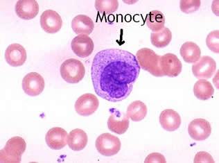
- MonocytesMonocytes comprise 3-8% of all WBCs. They are the largest of the WBCs, 12-20 µm in diameter. The nucleus is indented or kidney shaped, not rounded, and surrounded by foamy cytoplasm. Their size is the most easily identifiable characteristic. Monocytes are slow to respond to infection but arrive in larger numbers. When stimulated they enlarge and differentiate into wandering macrophages that clean up cellular debris and microbes following infection.
Thrombocytes
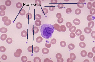
- PlateletsThrombocytes, or platelets, are numerous (150,000-400,000 platelets/mL blood) with a short 5-9 day life span. Platelets are not WBCs, they are small cell fragments (2-4 µm diameter) with many vesicles and no nucleus. They often appear as spots or “dirt” between the RBC’s. Platelets function to plug small holes in vessels and stop bleeding. The granules contain many factors involved in blood clotting, including clotting factors, platelet derived growth factor (PDGF), Ca++, ADP, ATP, Thromboxane A2, vasoconstrictors, and clot promoting enzymes.